
A nuclear stress test evaluates blood flow to the heart and helps diagnose heart conditions. When a patient undergoes this test, it helps a doctor observe how the heart functions during physical exertion compared to when it is at rest. The procedure provides detailed images of the heart, giving medical professionals a view of its performance under stress. Here is more information about how nuclear stress tests help diagnose heart conditions:
Measures Heart Function
Observing the heart’s response to stress is a primary objective of this test. This test shows how well blood is moving through the coronary arteries, and it helps determine if any blockages are present. If blood flow is restricted, the heart muscle may not receive enough oxygen, a condition that the test is designed to detect.
The test combines exercise with imaging technology. This approach enables a comprehensive evaluation of heart function, providing a clearer picture than exercise alone. Your doctor gathers information about the size of the heart’s chambers, its pumping function, and any muscle damage.
Monitors Patient During Exercise
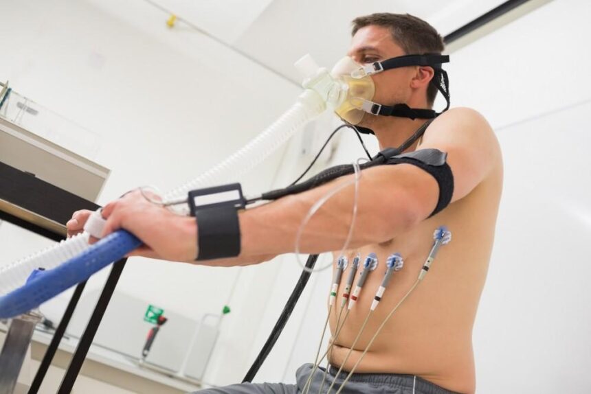
During the exercise portion of the test, your heart rate and blood pressure are continuously monitored. You may walk on a treadmill or pedal a stationary bike, and the intensity will gradually increase to make your heart work harder. If you are unable to exercise, doctors will administer a medication that simulates the effects of physical activity on your heart.
Your doctor may observe your electrocardiogram (ECG) throughout the test. A medical team records ECG patterns, along with any symptoms, such as chest discomfort or shortness of breath. This data, when combined with the imaging results, offers a detailed assessment of your heart’s condition.
The team administering the test is ready for any issues that might arise. The exercise continues until you reach a target heart rate, develop symptoms that prevent you from continuing, or show specific changes on the ECG. Your safety is the priority throughout the procedure.
Identifies Specific Details
The imaging part of a nuclear stress test yields specific details about your heart’s health. The test can identify the location and size of areas with poor blood flow, providing detailed information. When the heart is not getting enough blood, the images will show “cold spots,” which are areas where the radioactive tracer has not been absorbed.
Involves Radioactive Tracer
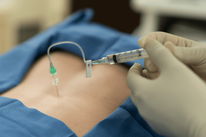
This test requires the use of a small, safe amount of radioactive material, called a tracer. The tracer is injected into your bloodstream through an intravenous (IV) line, and it travels to your heart muscle. A special camera, known as a gamma camera, detects the radiation from the tracer. It produces images of your heart.
Two sets of images are typically taken:
- One set is captured while your heart is at rest.
- Another set is taken after your heart has been stressed.
By comparing these two sets of images, a doctor can see how blood flow changes between rest and exertion. The amount of radioactive material doctors use is small, and it is gone from your body within a day or two.
Schedule a Nuclear Stress Test
A nuclear stress test provides a detailed look at the blood flow to your heart. While the procedure involves both exercise and imaging, it gives your doctor a comprehensive view of your heart’s function. The results from this test help guide decisions about your health. If you and your doctor have discussed this procedure, contact a clinic today to schedule your appointment.