

Magnetic resonance imaging (MRI) is a diagnostic tool used in healthcare settings. It utilizes strong magnetic fields and radio waves to create detailed images of the organs and tissues within the body. Here is more information on MRI scans, why they are conducted, what the process involves, their benefits, and how to prepare:
What Is an MRI Scan?
Magnetic resonance imaging is a non-invasive medical imaging technique used in radiology. The scanner itself typically resembles a large tube with a table in the middle, allowing the patient to slide inside. Unlike X-rays or CT scans, an MRI scan does not use ionizing radiation, and it relies on magnetic fields. This technology allows physicians to view the body’s internal structures with high precision.
The machine works by realigning water molecules in the body. Radio waves manipulate these protons, and they emit faint signals that are picked up by the receiver. A computer processes these signals to construct cross-sectional images, which can be viewed from different angles. This method produces three-dimensional representations of soft tissues and bones.
Why Is It Conducted?
Doctors recommend MRI scans to diagnose or monitor a wide variety of medical conditions. It serves as a primary tool for examining the brain and spinal cord, and it helps assess complex joint injuries. The scan detects tumors, cysts, and other structural abnormalities in various parts of the anatomy. Physicians rely on these detailed images to formulate accurate diagnoses and treatment plans.
The technology is also applicable to the heart and blood vessels. It assesses the structure of the heart’s chambers, and it identifies blockages or inflammation in the vessels. Scans of the liver, kidneys, and other abdominal organs provide data on function and health.
What Does the Process Involve?
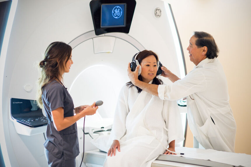
The patient lies on a movable table that slides into the center of the cylindrical magnet. The machine creates a strong magnetic field around the body, and radio waves are directed at the specific area of interest. You remain still during the scan, or the resulting images may appear blurry and unusable. The technologist communicates with the patient throughout the procedure.
Loud tapping or thumping noises occur rhythmically while the machine captures images. Earplugs or headphones are provided to dampen mechanical noise, and music may be played to help the patient feel more comfortable. The scan typically lasts 20 minutes, depending on the body part being examined. Some examinations require the use of a contrast material.
What Are the Benefits?
MRI scans provide clear images of soft-tissue structures. This level of detail enables early detection of disease, and it assists in monitoring the effectiveness of ongoing treatments. The procedure is non-invasive, and it doesn’t use ionizing radiation. The detailed images support surgical planning, as surgeons use the data to map out procedures before entering the operating room.
How Can You Prepare?
Patients remove all metal objects, such as jewelry, watches, and hairpins, before entering the scanning room. The strong magnetic field attracts metal, and these items can distort the final images. Inform the technician if you have any metal implants, pacemakers, or shrapnel in your body. Follow the instructions provided by your healthcare provider regarding food and routine medication.
Prepare for Your Scan Today
Understanding the MRI process helps facilitate a smooth procedure. Proper preparation contributes to high-quality images, and it enables the medical team to obtain the necessary diagnostic information. Consult your physician if you have additional questions regarding your specific scan instructions. Schedule your appointment and arrive ready for a successful imaging experience.