

A stress test evaluates how the heart responds to physical activity. By increasing the heart’s workload, this test reveals potential patterns in blood flow or irregularities that may not surface when at rest. It enables cardiologists to detect concerns such as coronary artery disease. Here’s more information about stress testing and how it can help diagnose heart conditions:
Understanding the Purpose of Stress Testing
The primary purpose of stress testing is to observe the heart’s reaction to exertion; this insight helps assess whether the heart muscle receives the necessary blood flow when under increased demand. Some patients may find that exertional symptoms, such as shortness of breath or dizziness when walking, lead them to seek testing. Others undergo stress evaluation due to their health history.
Stress testing provides data that is useful for determining safe levels of activity and assessing the heart’s capacity to handle daily physical demands. By recording specific measurements, such as heart rate, blood pressure, and electrical activity, the test offers a systematic way to examine heart performance during controlled activities. This process is helpful for individuals and their healthcare providers when developing personalized care plans.
Describing the Stress Testing Procedure
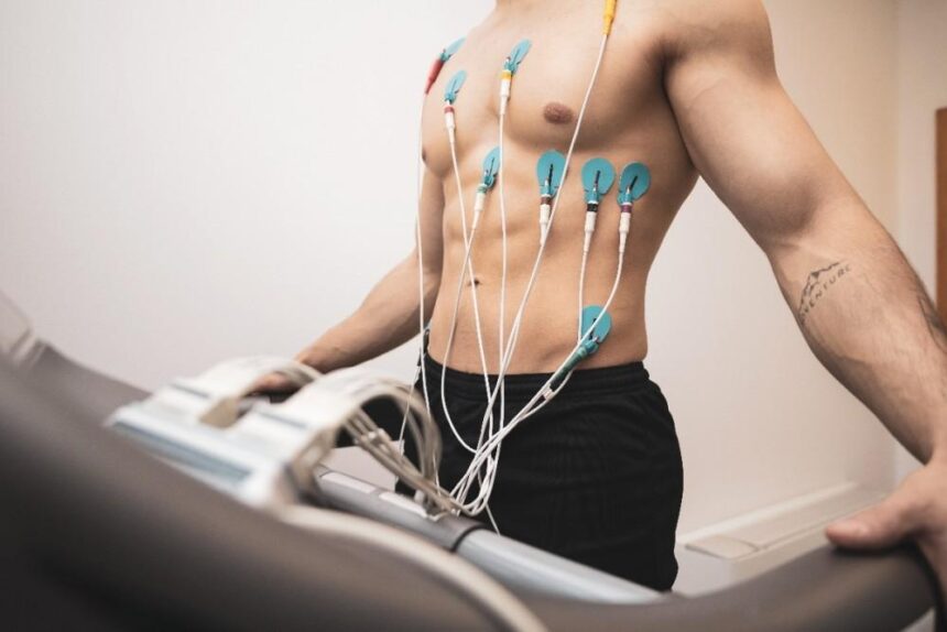
A stress testing procedure can involve exercise modalities like treadmills or stationary bicycles. Individuals undergoing the test have electrodes attached to their bodies, allowing continuous monitoring of heart rhythm through an electrocardiogram (ECG). A cuff may also be used to measure blood pressure throughout the session.
Exercise begins at a modest intensity and increases at set intervals. The session continues until a target heart rate is reached, symptoms arise that require a pause, or notable changes appear in the heart’s measurements. For individuals unable to exercise due to physical limitations, medications can be used as an alternative. These drugs simulate the effects of physical activity, prompting the heart to respond as it would during exercise.
Incorporating Imaging Into Stress Testing
Some stress tests utilize imaging to gain further insight into heart function; a cardiac positron emission tomography (PET) scanner is one option. This technique provides detailed images of blood flow and identifies areas of the heart that may not be receiving adequate oxygen. Combining imaging with exercise or medication-induced stress allows cardiologists to make more precise diagnoses and tailor treatment plans effectively.
Imaging-based stress tests include echocardiogram stress tests and nuclear stress tests. An echocardiogram stress test uses ultrasound to capture images of the heart before and after exercise-induced stress. These images display the heart’s structure, chamber size, wall motion, and valve function. Healthcare providers may examine these results to identify regions of the heart muscle that respond differently under stress.
A nuclear stress test involves the introduction of a small amount of radioactive tracer into the bloodstream. Special cameras create images displaying blood flow to the heart muscle at rest and after stress. This methodology helps visualize the relative blood supply in various areas of the heart, and it can identify potential blockages in the coronary arteries.
Meet With a Cardiologist
Stress testing is a valuable tool for detecting underlying heart conditions, such as arrhythmias or reduced blood flow. By evaluating how the heart responds to controlled physical or medication-induced stress, cardiologists can identify potential risks and determine safe activity levels. The test also enables them to develop personalized treatment plans and support long-term cardiovascular health. Contact a qualified cardiologist today to receive guidance tailored to your individual circumstances.